ACHALASIA CARDIA
ACHALASIA CARDIA
INTRODUCTION
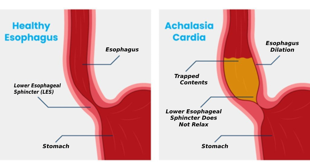
Oesophagus or food pipe (अन्ननलिका) is a muscular tube which connects the mouth to the stomach. When food or drink is swallowed, muscles in the food pipe contract and relax to push food into the stomach. This is known as peristalsis. At the bottom of the oesophagus and near its junction with the stomach is a valve called the lower oesophageal sphincter (LES).
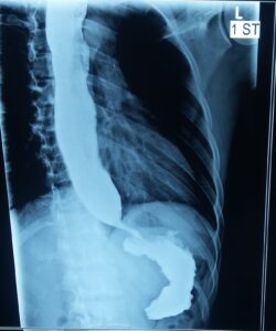
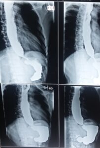
When you swallow, the lower oesophageal sphincter opens to allow food and drink into the stomach. In achalasia, the muscles in the oesophagus do not contract correctly, and the ring of muscle at the bottom end (LES) can fail to open properly, or does not open at all. As a result, the oesophagus loses its ability to transfer food and drink into the stomach. This is a problem which affects the nerves and muscles of the oesophagus and LES.
CAUSES:
Achalasia is a rare condition that affects around one to two in every 100,000 people. It is usually diagnosed in adults but may occur in children. In most patients, there is no specific cause, and there is nothing a person can do to stop it.
In some patients, however it may be linked to a viral infection. It is also thought to be associated with having an autoimmune condition, where the body’s immune system attacks healthy cells, tissue, and organs.
In rare cases, it’s possible that achalasia is caused by genetic disorder.
Achalasia is thought to happen when the nerves in the oesophagus become damaged and stop working properly, which is why the muscles and ring of muscle at the lower end do not work.
PRESENTATION:
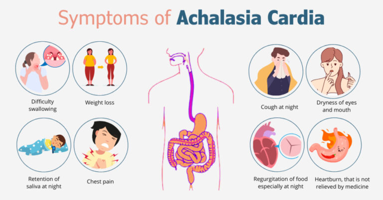
- Difficulty in swallowing: Patients with achalasia have difficulty passing food and drink into the stomach. Initially large and solid pieces of food such as non-veg and bread, naan, etc. may be difficult to pass but later drinking water may become difficult.
- Chest pain
- Coughing bouts or choking
- Regurgitation or vomiting of undigested food and drink.
- Heartburn
- Repeated chest infections
- Drooling of vomit or saliva
- Gradual but significant weight loss
TYPES:
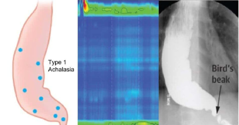
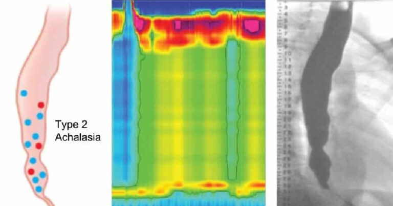
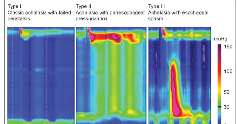
There are three major types of achalasia
-
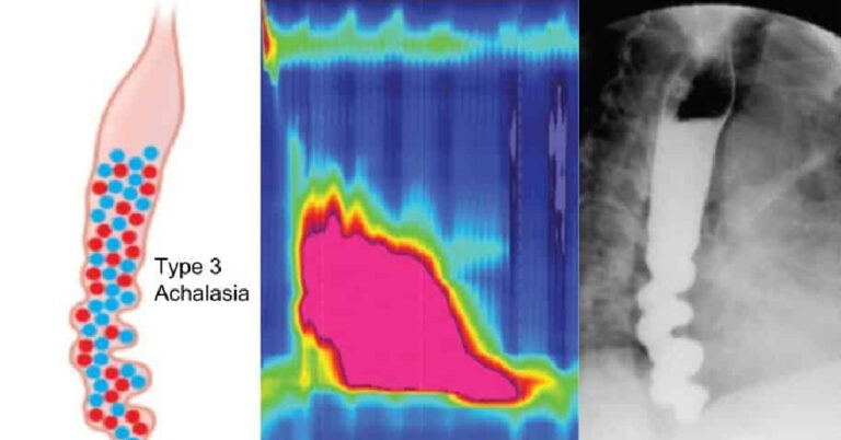
Type III achalasia
Type III, sometimes called spastic achalasia because there are abnormal contractions at the bottom of the oesophagus where it meets the stomach. This is the most severe type of achalasia. The contractions can cause chest pain that can awaken a person from sleep and imitate the symptoms of a heart attack.
Investigations:
-
Endoscopy (Gastroscopy)
This procedure involves passing a long thin telescope through the mouth and into the oesophagus and stomach. This gives a clear view of the lining of the oesophagus and stomach. Doctor can also take biopsy samples if needed.
-
Oesophageal manometry
This is the most useful test for achalasia. A thin plastic tube is passed via the nose, down the oesophagus and into the stomach. You will then be asked to take swallows of water, and then some rice, whilst recordings are made. The tube measures the pressure along your food- pipe and in the lower oesophageal sphincter. Oesophageal manometry helps to identify the type of achalasia you have. This helps the doctors to decide which treatment is best for you.
How is achalasia treated?
Depending on the type , there are various ways to treat Achalasia. All treatments aim to open up the passage between the oesophagus and the stomach so that food and drink can pass through. It is important to note that achalasia should not be left untreated because it will never get better on its own. It will only become worse and more difficult to treat over time.
-
Medicine
Oral medicines, such as nifedipine (calcium channel blockers) or nitrates, can help to relax the muscles in your oesophagus. This makes swallowing easier and less painful for some people, although they do not work for everyone.
The effect is short lived, so medicine may be used to ease symptoms while you wait for a more permanent treatment. They may cause headaches.
-
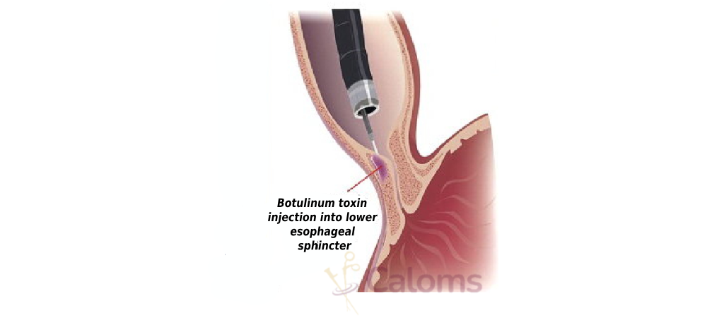
Botulinum toxin (Botox®)
This is a treatment in which Botox® is injected into the lower oesophageal sphincter during endoscopy. The Botox® paralyses the muscles in the lower oesophageal sphincter which allows food to pass into the stomach. It can be done as a day case procedure under light anaesthesia.
The effects, however may not be last long and the treatment may need to be repeated every 6 to 12 months. Therefore, Botox® is usually offered to patients who are not fit or cannot have any other sort of treatment.
-
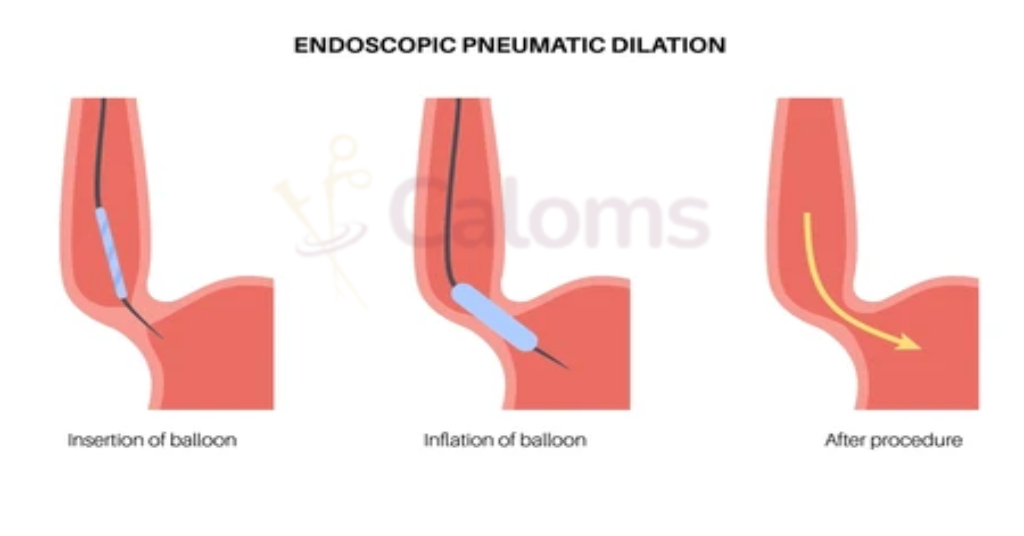
Balloon dilatation
The lower oesophageal sphincter is dilated or stretched with a balloon during an endoscopy and with the use of X-rays.
This treatment may require 2- 3 sittings, few weeks apart. The main complication is that there is a small risk (around 2% ) of a tear or perforation (a hole) in the food pipe which may require emergency surgery.
-
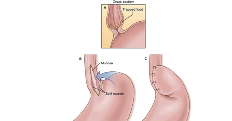
Surgery
During surgery, muscles in the lower oesophageal sphincter are cut in an operation called a Heller myotomy. Normally this is performed with keyhole surgery (laparoscopy). To reduce the risk of heartburn and severe acid reflux, an anti-reflux procedure (Fundoplication) is normally done at the same time. The procedure is performed under general anaesthetic. It can permanently make swallowing easier.
-
Peroral endoscopic myotomy (POEM)
During surgery, muscles in the lower oesophageal sphincter are cut in an operation called a Heller myotomy. Normally this is performed with keyhole surgery (laparoscopy). To reduce the risk of heartburn and severe acid reflux, an anti-reflux procedure (Fundoplication) is normally done at the same time. The procedure is performed under general anaesthetic. It can permanently make swallowing easier.
What is the recovery like after surgery?
- By evening patients are able to walk and can have fluids by mouth.
- Most of patients go home after 24 hours, however some may have to stay for a day extra depending on recovery and social circumstances.
- You can return to non-strenuous activity (going to office) within a couple of days, but you must avoid strenuous activity for six weeks.
- You should not drive for 2 weeks however you can travel by cab.
What is the dietary advice following surgery?
- There are restrictions to food intake for the first few weeks after the operation.
- You should only drink water for the rest of the day after the operation.
- Our dietician will formulate a diet plan for next four weeks depending on your social and religious beliefs.
- Some patients become constipated and need to take laxatives if their bowels have not opened.
- If you have any difficulties with certain foods discuss this with your consultant or dietitian. At your 4 week follow-up appointment with your consultant/ dietician you may start a normal diet.
Cost of ACHALASIA CARDIA
Cost depends on lots of factors such as
- Bed category
- Type of surgery
- Open or laparoscopic repair
- Choice of hospital
- Preexisting medical conditions such as diabetes, angina which may prolong your hospital stay or need critical monitoring. Following your first meeting with the doctor, we would be able to give you an approximate estimate.

